

A bone age study helps doctors estimate the maturity of a child’s skeletal system. It’s usually done by taking a single X-ray of the left wrist, hand, and fingers. It is a safe and painless procedure that uses a small amount of radiation. The bones on the X-ray image are compared with X-rays images in a standard atlas of bone development, which is based on data from large numbers of other kids of the same gender and age. The bone age is measured in years.
A child’s bones, such as those in the fingers and wrist, contain “growing zones” at both ends called growth plates. These plates consist of special cells responsible for the bones’ growth in length. Growth plates are easy to spot on an X-ray because they’re softer and contain less mineral, making them appear darker on an X-ray image than the rest of the bone.
As kids grow, growth plates change in appearance on the X-ray images and become thinner, eventually disappearing (this is what is called “closed growth plates”). Because they look different at each age, a doctor can assign a bone age based on the appearance of the bones and growth plates. A child’s bone age (also called the skeletal age) is assigned by determining which of the standard X-ray images in the atlas most closely match the appearance of the child’s bones on the X-ray.
A difference between a child’s bone age and his or her chronological age might indicate a growth problem. But such differences don’t always mean there’s a problem, because perfectly healthy kids can have bone ages that differ from their chronological ages.

Why It’s Done
The bone age study can help evaluate how fast or slowly a child’s skeleton is maturing, which can help doctors diagnose conditions that delay or accelerate physical growth and development. This test is usually ordered by pediatricians or pediatric endocrinologists.
Bone age can be used to predict:
- how much time a child will be growing
- when a child will enter puberty
- what the child’s ultimate height will be
The test is also used to monitor progress and guide treatment of kids with conditions that affect growth, including:
- diseases that affect the levels of hormones involved in growth, such as growth hormone deficiency, hypothyroidism, precocious puberty, and adrenal gland disorders
- genetic growth disorders, such as Turner syndrome (TS)
- orthopedic or orthodontic problems in which the timing and type of treatment (surgery, bracing, etc.) must be guided by the child’s predicted growth

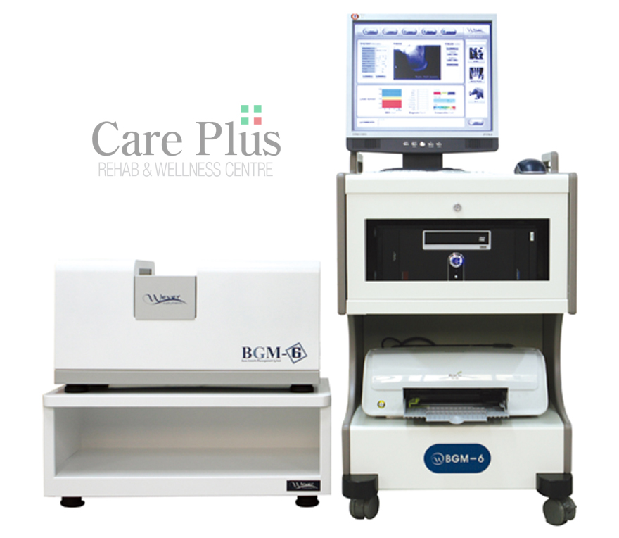
Children’s bone density measurement(a growth clinic aid program)
1) Providing X-ray images of upgraded resolution
- Calculation of the length and area of growth plates and bones
- Internally provided with image magnification and rotation functions
2) Providing a graph to compare height and weight with standard values at the relevant age
3) Measuring children’s calcaneus density(mg/cm2) and then indicating it on the graph by age
4) Providing 3 kinds of age concepts that are mentioned in the pediatric growth theory
- Chronological age : Medical age
- Height age(50% percentile) : Growth index age in terms of height using statistical data
- Bone age : Analyzed with 3 kinds of theories
① Calculating bone age with RUS scores among TW3 methods and expressing it with graphs
② Calculating bone age with Carpal scores among TW3 methods and expressing it with graphs
③ Calculating bone age in a G-P Atlas method
5) Internally provided with the MRUS function to minimize errors in the bone age value that can differ depending on readers(lessening the bone age reversal phenomenon due to reading bone age for a short period)
6) Providing prediction index for adult’s height by 4 kinds of theories
- AHP : TW3(RUS score) method of prediction index for adult’s height using bone age concepts
- SHP : Prediction index for adult’s height using statistical techniques
- GHP : Prediction index for adult’s genetic height
- KHP : Prediction index for adult’s height using the Khamis-Roche method (simultaneously considering a genetic concept of current growth status)
- AVG : Indication of the average value of adult’s height measured by AHP, SHP, GHP, and KHP
7) Internally provided with a program to predict growth periods
: EGP(suspension of growth), EMP(appearance of menarche), EVP(secondary rapid growth period)
8) Providing a graph of comparison with standard values of BMI(fatness index) at the relevant age
9) Follow-up(record control & comparison between pre and post-clinic) expression Expression of results of comparing and analyzing examination contents over many times for a specific patient in order to have progress status of growth clinics compared and judged
- Graphic expression of changes in bone age and height age pursuant to age
- Graphic expression of changes in adult’s predicted height(AHP, SHP, GHP, KHP, AVG)
- Expression of patient’s growth speed in cm/year
- Graphic expression of changes in height and weight by age
- Indication of years when one can grow in the future(i.e., indication of age when growth stops)